
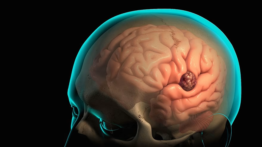
Glioblastoma — classified by the World Health Organization as a Grade IV, IDH-wildtype astrocytoma since the 2021 CNS tumor update — accounts for roughly 49 percent of all malignant brain cancers diagnosed in the United States each year, yet strikes only about 3.19 people per 100,000. That gap between dominant among its category and rare in absolute terms shapes the entire experience of the disease: most people have never encountered it, but those who do face some of the starkest survival numbers in oncology. Left entirely untreated, the prognosis for untreated glioblastoma is a median survival of approximately three months from diagnosis. With the best currently available treatment — surgery, radiotherapy, and chemotherapy — that figure climbs to roughly 14 to 16 months. Understanding why the gap is not larger requires looking closely at what glioblastoma actually is, and why it resists every tool medicine brings to bear.
What is glioblastoma and how does it differ from other brain tumors?
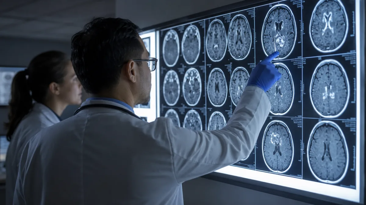
Glioblastoma arises from astrocytes, the star-shaped support cells woven throughout the brain's architecture. Unlike neurons, which cannot divide after development, astrocytes retain some capacity for proliferation — and in GBM, that capacity is catastrophically unregulated. The 2021 WHO Classification of Central Nervous System Tumors redrew the boundaries meaningfully: what was once called "glioblastoma, IDH-mutant" is now reclassified as Astrocytoma, IDH-mutant, Grade 4, a biologically distinct entity with a considerably better outlook. True glioblastoma, under the new schema, is IDH-wildtype by definition, and requires at least one of the following molecular fingerprints: EGFR gene amplification, TERT promoter mutation, or a combined gain of chromosome 7 alongside loss of chromosome 10.
These molecular features matter clinically, not just diagnostically. EGFR amplification — present in roughly 40 percent of GBM cases, according to the American Brain Tumor Association — drives aggressive cell proliferation through persistent growth-signal cascades. TERT promoter mutations extend tumor cell lifespan by reactivating telomerase, the enzyme that prevents chromosomal shortening during division. The net effect is a tumor that grows fast, thinks little about dying, and spreads into surrounding tissue through finger-like projections that make clean surgical margins essentially impossible.
A second molecular marker, MGMT promoter methylation, is not part of the WHO diagnostic criteria but carries enormous prognostic and predictive weight. When the MGMT gene's promoter is methylated (chemically silenced), the tumor loses its primary DNA-repair enzyme — the one that would normally undo the damage caused by chemotherapy drugs like temozolomide. Around 40 to 50 percent of GBM patients have MGMT-methylated tumors, and this group consistently shows better responses to temozolomide and longer overall survival. For the remaining patients, whose MGMT gene remains active and repairs chemotherapy-induced damage, the practical benefit of the drug is substantially diminished.
Glioblastoma most often forms in the frontal and temporal lobes, though any supratentorial region can be affected. It can cross to the opposite hemisphere via the corpus callosum — a growth pattern sometimes called a "butterfly" lesion — and rarely spreads outside the central nervous system entirely.
What are the symptoms of glioblastoma?
Symptoms vary depending on where within the brain the tumor is growing, and many patients experience only vague complaints for weeks before a diagnosis is made. The most characteristic headache pattern is one that worsens in the morning or when lying down — positions that increase intracranial pressure — and does not respond reliably to over-the-counter pain relief. This distinguishes it, at least in retrospect, from tension headache or migraine, though GBM headaches are frequently misattributed to those conditions early on.
Seizures affect a significant proportion of patients at presentation, particularly when the tumor involves the cortex. These are not always the convulsive, full-body events most people associate with epilepsy. Focal seizures from a frontal lobe GBM might present as uncontrollable twitching in one hand; temporal lobe involvement can trigger unusual smells, a rising sense of dread, or brief episodes of unresponsiveness. A first seizure in an adult without prior epilepsy history is a medical red flag requiring urgent brain imaging.
Focal neurological deficits depend on location. Tumors in motor cortex produce weakness or paralysis on the opposite side of the body. Left-hemisphere lesions near Broca's or Wernicke's areas cause expressive or receptive aphasia — the inability to produce or understand speech, respectively. Occipital involvement can cause visual field defects, while deep frontal lesions often manifest as personality change, disinhibition, or loss of executive function that families notice long before the patient does. Cognitive changes — memory loss, slowed thinking, difficulty with planning — are common regardless of location, partly from the tumor itself and partly from the edema that surrounds it.
Who is most likely to develop glioblastoma?
According to the CBTRUS Statistical Report, the incidence of GBM is 3.19 per 100,000 persons in the United States, with a median age at diagnosis of 64 years. Incidence is approximately 1.6 times higher in males than in females — a disparity that has been observed consistently across decades and registries but remains incompletely explained. It is also roughly twice as common in white populations compared to Black populations, with lower rates in Asian and American Indian groups.
Age is the most powerful risk factor. Incidence rises steeply after age 50 and peaks between 75 and 84 years. GBM can and does occur in younger adults and, rarely, in children — but the overwhelming majority of diagnoses land in the sixth decade of life and beyond. Among children, GBM represents fewer than 10 percent of pediatric brain tumor cases, and when it does occur in young patients, molecular differences often make the biology somewhat more favorable than in older adults.
No modifiable lifestyle risk factor has been convincingly established. Ionizing radiation exposure — particularly prior therapeutic radiation to the head — is the one confirmed environmental risk. Claims linking mobile phone use or dietary factors to GBM have not survived rigorous epidemiological scrutiny. Most patients who receive the diagnosis have no identifiable predisposing factor, which offers little comfort but does mean that GBM is not something that could have been prevented through different choices.
What is the prognosis for untreated glioblastoma?
The data here are unambiguous and should be understood directly. Without any treatment, the median survival for a patient with newly diagnosed GBM is approximately three months, as documented in multiple NCBI and peer-reviewed sources. "Median" means half of untreated patients die within that window; a smaller number may survive marginally longer, but very few reach six months. The tumor's doubling time is rapid, and the mechanisms driving death — herniation from mass effect, loss of brain function from infiltration, hemorrhage into necrotic tumor tissue — proceed without interruption when no therapy is in place.
This three-month figure is sometimes cited alongside a caution: it comes largely from historical series and from subgroups of clinical trial populations who declined or were ineligible for treatment, so it carries some methodological caveats. But across different datasets and time periods, the number has been consistently grim. Even in registry data that includes patients who received supportive care only, median survival rarely exceeds six months.
Some patients and families encounter this prognosis and ask whether doing nothing — avoiding the burdens of treatment — might be a legitimate choice. That is a question only the patient can answer, in consultation with their care team. What the evidence supports is that no treatment produces reliably the worst survival outcome, and that the decision to forego active therapy is meaningfully different from the decision to pursue comfort-focused palliative care alongside or instead of aggressive intervention. These are not the same thing.
How does standard treatment affect glioblastoma survival?
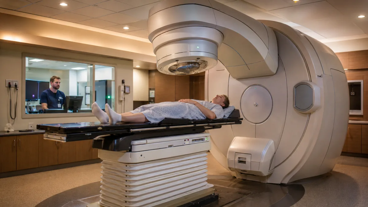
The current standard of care for newly diagnosed GBM was established by a landmark trial published in the New England Journal of Medicine in 2005 by Roger Stupp and colleagues. The Stupp protocol randomized 573 patients across 85 centers to receive either radiotherapy alone or radiotherapy combined with the oral chemotherapy agent temozolomide, given both concurrently with radiation and for six subsequent monthly cycles. Results were decisive: the combination arm achieved a median overall survival of 14.6 months compared to 12.1 months for radiation alone, and the two-year survival rate was 26.5 percent versus 10.4 percent. Long-term follow-up showed that approximately 9.8 percent of the temozolomide group survived five years — a figure that would have seemed impossible before the trial, though it also underscores how few patients reach that milestone.
The benefit of temozolomide was not uniform. Patients with MGMT-methylated tumors drove much of the survival advantage, with two-year survival rates in that subgroup reaching above 40 percent in some analyses. For those with unmethylated MGMT, the drug still contributes to treatment but with considerably reduced effect. Identifying MGMT methylation status is now a standard part of molecular workup at diagnosis.
In 2015, tumor treating fields (TTFields), marketed as Optune, received FDA approval as an addition to maintenance temozolomide for newly diagnosed GBM. The phase III EF-14 trial, which enrolled 695 patients, showed that adding TTFields — a device worn on the scalp that delivers low-intensity alternating electric fields to disrupt tumor cell division — extended median survival from 19.8 months to 24.5 months when added to temozolomide. Five-year survival in the TTFields arm reached 13 percent. The device is cumbersome and requires patients to wear electrode arrays on the shaved scalp for at least 18 hours per day, but it does not appear to impair cognitive function or quality of life in the way that some systemic therapies do.
Surgery remains the first step whenever safely possible, serving three purposes: it establishes a tissue diagnosis, reduces tumor bulk and intracranial pressure, and creates conditions under which radiation and chemotherapy operate more effectively. Achieving a gross total resection — removing all visible tumor on MRI — is associated with longer survival, but it does not cure the disease. Infiltrative cells beyond the resection margin survive and will drive recurrence.
What makes glioblastoma so difficult to treat?
Three biological features of GBM conspire against effective therapy, and understanding them clarifies why incremental advances in treatment have not translated into the dramatic survival gains seen in some other cancers.
The first is the blood-brain barrier. The tight junctions between endothelial cells lining the brain's capillaries form a selective filter that evolved to protect neural tissue from circulating toxins. Most chemotherapy agents are too large or too hydrophilic to cross it in effective concentrations. Temozolomide is an exception — it is small, lipophilic, and capable of reaching the brain — which is precisely why it became the backbone of GBM chemotherapy. Bevacizumab, a monoclonal antibody targeting the vascular growth factor VEGF, was approved for recurrent GBM partly because anti-angiogenic therapy disrupts the leaky tumor vasculature; however, it has consistently improved progression-free survival without extending overall survival, suggesting that the tumor adapts rather than surrenders.
The second obstacle is intratumoral heterogeneity. A single GBM contains multiple genetically distinct cell populations, each potentially carrying different mutations and different sensitivities to therapy. A treatment effective against the dominant clone may leave resistant subclones untouched; under the selective pressure of therapy, those subclones proliferate and the tumor that recurs is biologically different from the one that was treated. This is why GBM almost universally recurs, and why second-line treatment outcomes are substantially worse than first-line results.
The third factor is the infiltrative growth pattern. GBM cells spread along white matter tracts and perivascular spaces far beyond the contrast-enhancing mass visible on MRI. Studies using postmortem tissue analysis and advanced imaging have identified viable tumor cells centimeters beyond the surgical resection margin. Approximately 90 percent of recurrences occur within two centimeters of the original tumor bed, but the infiltrative cells that seed those recurrences are present from diagnosis, beyond the reach of any scalpel and protected by a partially intact blood-brain barrier in the peritumoral zone.
What newer treatments are being researched for glioblastoma?

Immunotherapy has transformed outcomes in several solid tumors, and intensive research has sought to apply similar approaches to GBM — with more limited success so far, though the field is moving.
Checkpoint inhibitors, which release the brakes on T-cell activity, have shown strong results in cancers like melanoma and lung cancer. In GBM, multiple phase III trials of checkpoint inhibitors including nivolumab have not met their primary survival endpoints, likely because the brain's immune microenvironment is profoundly immunosuppressive, limiting T-cell trafficking and activation. Checkpoint inhibitors comprised about 26 percent of GBM immunotherapy trials as of 2025, and combination strategies — pairing them with other agents or TTFields — remain under investigation.
CAR-T cell therapy has produced some of the most watched early results. A bivalent CAR-T construct targeting both IL13Rα2 and EGFRvIII — proteins highly expressed on GBM cells — was evaluated in 18 patients as of a June 2025 report, with findings emphasizing both the promise of locoregional intracranial delivery and the challenges of scaling this approach. Responses have been observed, but durable remissions remain elusive and the field is still early.
Cancer vaccines using patient-derived tumor antigens aim to train the immune system to recognize and attack GBM cells. A phase I/II trial of SL-701, a glioma-associated antigen vaccine, enrolled 74 patients with recurrent GBM in 2024. Results are pending long-term analysis but have informed the design of subsequent combination trials.
Preclinical work on nanoparticle drug delivery systems — designed to carry chemotherapy agents across the blood-brain barrier — has shown survival improvements in animal models, and Mayo Clinic announced a dual-drug nanotherapy approach that crossed the blood-brain barrier in preclinical GBM models. Translation to human trials is underway but will take years to yield definitive clinical evidence.
What is palliative care for glioblastoma patients?
Palliative care is not the same as giving up. For GBM patients, it refers to an integrated layer of medical, psychological, and social support that runs alongside — and eventually may replace — active tumor-directed treatment. Its goals are to reduce symptom burden, preserve cognitive and functional independence for as long as possible, and ensure that patients and families have the information and support to make decisions that reflect their own values.
Corticosteroids, most commonly dexamethasone, are used in up to 87 percent of high-grade glioma patients to control cerebral edema. Dexamethasone works by reducing the permeability of tumor blood vessels and dampening the inflammatory response around the tumor, which can dramatically reduce headache and improve neurological function within days. The trade-off is a significant side-effect profile at sustained doses — including diabetes, immune suppression, adrenal insufficiency, and a painful myopathy — so the principle is to use the lowest effective dose and taper as quickly as symptoms allow.
Seizure control, fatigue management, cognitive rehabilitation, and psychological support for both patients and caregivers form the core of palliative practice in GBM. As the disease progresses, early integration of hospice planning — including conversations about advance directives, goals of care, and what "good days" look like — has been shown to reduce the likelihood of aggressive, burdensome interventions in the final weeks that do not improve quality of life. Hospitalization in the last month of life, and particularly intensive care admission, is associated with worse quality of life outcomes without survival benefit.
Palliative care teams at comprehensive cancer centers specialize in navigating these questions, and patients do not have to wait until tumor-directed options are exhausted to request a palliative consult. Early integration is now recommended by major oncology organizations as part of standard GBM management.
This article is for general informational purposes only and does not constitute medical advice. Individuals facing a glioblastoma diagnosis should work closely with a neuro-oncology team to understand treatment options specific to their situation.
For families and patients seeking deeper information, the American Brain Tumor Association's GBM resource page, the National Cancer Institute's adult brain treatment guide, and the Mayo Clinic's glioblastoma overview provide regularly updated, clinically reviewed information. For those seeking to understand what causes gliomas at a molecular level, or who want to learn about metastatic bone cancer symptoms in the context of cancer spreading to secondary sites, additional resources are available on this site.
Frequently Asked Questions
How long can someone live with untreated glioblastoma?
Multiple peer-reviewed studies report a median survival of approximately three months for patients with GBM who receive no tumor-directed treatment. A small number of patients survive somewhat longer, but the disease progresses rapidly without intervention, and survival beyond six months without treatment is uncommon. Median survival refers to the midpoint — half of patients survive less than this figure, half survive more.
What is the best-case survival for GBM with treatment?
The Stupp protocol — surgery followed by concurrent temozolomide and radiotherapy, then adjuvant temozolomide — produces a median overall survival of 14.6 months. Adding tumor treating fields (Optune) extends median survival to approximately 24.5 months in trials. A subset of patients, particularly those with MGMT-methylated tumors, reach the five-year mark — around 9 to 13 percent depending on the treatment combination and the trial population. These figures represent group medians, not predictions for any individual patient.
What is MGMT methylation and why does it matter?
MGMT is a DNA repair enzyme. When the gene that codes for it has its promoter region chemically silenced — a process called methylation — the tumor cannot efficiently repair the DNA damage caused by temozolomide. This makes the chemotherapy more effective. Roughly 40 to 50 percent of GBM patients have MGMT-methylated tumors; they tend to live longer and respond more strongly to temozolomide than those whose MGMT gene is active.
Why does glioblastoma almost always come back?
GBM grows by sending infiltrative cells along white matter tracts and perivascular spaces into surrounding brain tissue. Imaging identifies the enhancing tumor mass, but individual cells migrate centimeters beyond the visible margin long before surgery. These cells survive resection and, protected by a partially intact blood-brain barrier in the peritumoral zone, are often out of reach of chemotherapy at effective concentrations. Around 90 percent of GBM recurrences appear within two centimeters of the original tumor site, driven by these surviving cells.
Is glioblastoma hereditary?
The vast majority of GBM cases have no hereditary cause. Rare inherited syndromes — including Li-Fraumeni syndrome, Lynch syndrome, neurofibromatosis, and Turcot syndrome — are associated with increased risk, but collectively account for a very small fraction of cases. Most patients who receive a GBM diagnosis have no family history of the disease and no identifiable genetic predisposing factor.
Are there clinical trials for glioblastoma patients?
Yes, and neuro-oncology specialists typically encourage participation given the limitations of current standard therapy. Active trials are exploring CAR-T cell therapies, next-generation checkpoint inhibitor combinations, cancer vaccines, nanoparticle drug delivery to overcome the blood-brain barrier, and novel combinations of tumor treating fields with immunotherapy. The National Cancer Institute maintains a searchable database of open trials at cancer.gov, and most comprehensive cancer centers have dedicated neuro-oncology trial programs.

Linda Fehrman
Edits general wellness and relationship explainers. Health material is educational, avoids diagnosis and links to health-authority guidance.




Leave a reply
Replying to