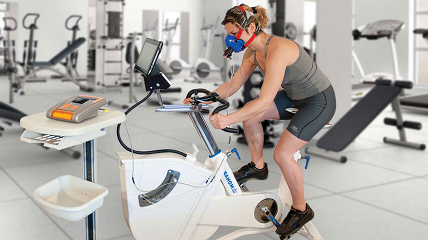
The Maximal Exercise Test to Determine Maximal Heart Rate
Exercise testing to determine an individual’s maximal heart rate is used for evaluating the body’s (particularly the heart’s) response to stress. As exercise is the body’s most common physiological stress, it is a practical test to assess flow of blood and function.
An electrocardiograph is used to collect data on the heart, producing a document known as an electrocardiogram, or EKG. A patient is typically exercised to dangerous levels under observation, to a level of 85% of the predicted maximal heart rate for their age, or 85 percent of 220 minus their age.

Exercise Physiology
To understand maximal heart rate during exercise, it is important to consider some basics of exercise physiology and testing. During exercise, muscle cells in the heart known as cardiac myocytes pump harder and faster, and this increases heart muscle oxygen demand. If the heart is not receiving enough oxygenation, this may cause cell death and myocardial infarction.
It is possible to estimate this demand by multiplying heart rate times systolic blood pressure (the higher of the two numbers), and in patients with low demand, we frequently see future myocardial ischemia and infarction. It is possible to detect dying and dead heart cells using an EKG.
Methodology
A stress test is usually performed using physical means, typically with a patient walking and then running up an inclined treadmill to induce exercise-based stress. However, in patients with back, joint or muscle problems or with heart or lung diseases, a pharmocologic stress test may be performed. In this test, drugs are administered to simulate the exercise response, typically adenosine, dipyridamole, regadenoson or dobutamine.
It is important for patients to avoid oral nitrates (including nitroglycerin) or caffeine before a pharmocological stress test.
Diagnosis
The EKG is used in this scenario to look for damage to the heart, with various changes in the heartbeat complex indicating different issues. Common examples include ST segment changes indicating varying degrees of cell death or inverted, or peak T, waves suggesting electrolyte abnormalities. In the event that damage is seen or suggested by the EKG, patients are typically sent on for further study.
Typically, a similar test is performed in which exercise is used to stimulate the heart, but instead of looking at the outside with an EKG, Single Photon Emission Computed Tomography and other nuclear imaging scans can be used to directly observe blood uptake by the heart during exercise.
Treatment
If a maximal exercise treadmill test indicates problems with the heart at maximum heart rate, various interventions may be used. In particular, doctors are most concerned with unstable angina and the possibility of non-ST-segment elevation myocardial infarction (NSTEMI).
Treatment starts with anti-ischemic drugs such as beta blockers, nitrates, morphine, calcium channel antagonists, and ACE-inhibitors, the latter of which is not actually an anti-ischemic but has positive effects in preventing disease-based changes to the ventricles of the heart. More severe defects may warrant transplants, grafts or pacemaker or implantable cardiac defibrillator use.
You Might Also Like :: How to Lose 10 Pounds With a Sweat Suit

