
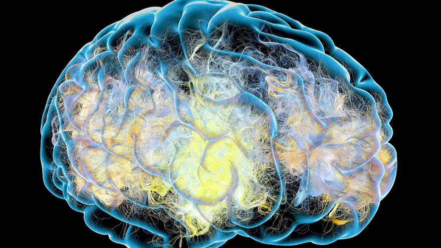
In 2017, the World Health Organization overhauled how brain tumors are classified, shifting glioma diagnosis from a purely visual assessment of tumor cells under a microscope to one grounded in molecular biology. That change, refined further in the 2021 WHO Classification of Tumors of the Central Nervous System, transformed how oncologists understand glioma causes, predict outcomes, and choose treatments. Gliomas are the most common primary malignant brain tumors in adults, and understanding what drives them — and what does not — matters enormously for anyone facing a diagnosis or trying to make sense of the research.
The focus keyword here is glioma causes, and the honest answer is that most cases arise without a clearly identifiable trigger. Two risk factors are well-established by evidence: prior exposure to ionizing radiation and certain inherited genetic syndromes. Everything else — from cell phone use to chemical exposures — sits in a more ambiguous space that deserves careful explanation rather than alarm or dismissal.
What is a glioma? Types and WHO classification
Gliomas originate in glial cells, the supportive cells that surround and maintain neurons throughout the brain and spinal cord. When glial cells accumulate DNA mutations that cause uncontrolled division and suppress normal cell death, they form a tumor mass. Unlike metastatic tumors — cancers that travel to the brain from elsewhere in the body — gliomas begin in brain tissue itself.
The 2021 WHO classification groups adult-type diffuse gliomas into three distinct entities, each defined by molecular markers as much as by appearance:
Astrocytoma, IDH-mutant (WHO Grades 2, 3, and 4): These tumors carry a mutation in the isocitrate dehydrogenase (IDH) gene and arise from astrocytes. They are graded 2 through 4 based on histological features including necrosis, microvascular proliferation, and specific chromosomal deletions. An IDH-mutant astrocytoma that reaches Grade 4 is distinct from glioblastoma, though both carry a poor prognosis at higher grades.
Oligodendroglioma, IDH-mutant and 1p/19q-codeleted (WHO Grades 2 and 3): Defined by both an IDH mutation and the simultaneous deletion of chromosomal arms 1p and 19q, oligodendrogliomas tend to grow more slowly and respond better to chemotherapy than most other gliomas. There is no Grade 4 oligodendroglioma under the 2021 system.
Glioblastoma, IDH-wildtype (WHO Grade 4): The most aggressive and most common malignant primary brain tumor. Glioblastoma multiforme (GBM) is by definition IDH-wildtype and always classified as Grade 4. It grows rapidly, invades surrounding brain tissue, and carries the worst prognosis of all adult glioma types.
This molecular shift matters practically: two tumors that look identical under a microscope may now be classified differently based on their IDH status, changing everything from prognosis to treatment strategy.
How common are gliomas?
According to the Central Brain Tumor Registry of the United States (CBTRUS), glioblastoma alone occurs at a rate of approximately 3.22 per 100,000 people per year, making it the most commonly diagnosed malignant primary brain tumor. Across all malignant primary brain and CNS tumors, the age-adjusted incidence rate was 6.89 per 100,000 for the period 2017–2021. Gliomas collectively account for roughly 80% of all malignant primary brain tumors.
Glioblastoma represents 13.9% of all primary brain tumors and 51.5% of all malignant ones. Peak incidence occurs between ages 45 and 65, and the disease is somewhat more common in men than in women and in white populations compared to Black populations in the United States, though the reasons for these demographic differences are not fully understood.
Confirmed risk factors for glioma
Despite decades of research, the list of confirmed glioma risk factors is short. The vast majority of cases arise in people with no obvious predisposing exposure. Two categories of risk are supported by strong evidence.
Ionizing radiation is the one environmental exposure with unambiguous scientific backing. People who have received therapeutic radiation to the head — for example, childhood leukemia treated with cranial radiotherapy — face a meaningfully elevated risk of developing a glioma years or decades later. The risk per Gray of radiation is highest in young children, particularly those treated before age five. Medical diagnostic radiation at typical doses is far lower risk, though high cumulative exposures remain a concern. This does not mean that routine CT scans or dental X-rays pose a significant glioma risk — the dose is not comparable — but it does establish the principle that ionizing radiation can cause DNA damage in glial cells that eventually leads to malignant transformation.
Age is a consistent risk modifier. While gliomas can occur at any age, and some types are more common in children, GBM predominantly strikes adults over 45. The accumulation of somatic mutations over time is the likely mechanism.
Beyond these two, no other environmental exposure has been confirmed as a glioma cause in large, well-controlled studies.
Genetic and hereditary risk factors
Most gliomas arise from mutations that develop during a person's lifetime — so-called somatic mutations — rather than from inherited genetic changes. That said, several rare hereditary syndromes substantially increase the risk of developing a glioma.
Neurofibromatosis type 1 (NF1) is caused by mutations in the NF1 tumor suppressor gene. It is among the most common hereditary conditions associated with brain tumors, particularly low-grade gliomas affecting the optic pathway in children.
Neurofibromatosis type 2 (NF2) involves mutations in the NF2 gene and is more strongly associated with meningiomas and schwannomas, but carries an elevated CNS tumor risk overall.
Li-Fraumeni syndrome, caused by germline mutations in the TP53 tumor suppressor gene, creates a broad cancer predisposition that includes brain tumors, soft tissue sarcomas, breast cancer, and leukemia. Brain tumors in Li-Fraumeni syndrome can appear in childhood.
Turcot syndrome, a variant of familial adenomatous polyposis or Lynch syndrome involving mutations in mismatch repair genes or the APC gene, is associated with both colorectal polyps and CNS tumors including glioblastoma.
Lynch syndrome, well-known for elevating colorectal cancer risk, also increases the risk of CNS tumors including gliomas through defects in mismatch repair genes such as MLH1, MSH2, MSH6, and PMS2.
A family history of glioma that does not fall into one of these recognized syndromes may still carry a modest inherited risk, though the specific genetic drivers are not fully characterized. Having a first-degree relative with glioma roughly doubles the risk, but because glioma is relatively rare, the absolute risk remains low.
At the molecular level, IDH mutations deserve attention because they illuminate a key mechanism of glioma development. In most Grade 2 and 3 gliomas and in secondary GBMs, the IDH1 or IDH2 enzyme is mutated to gain a new, abnormal function: rather than converting isocitrate to alpha-ketoglutarate, the mutated enzyme converts alpha-ketoglutarate into 2-hydroxyglutarate (2-HG) at more than 100 times the normal rate. This oncometabolite disrupts the function of alpha-ketoglutarate-dependent enzymes, including histone demethylases and DNA demethylases. The result is widespread abnormal methylation patterns across the genome — a state known as the CpG island methylator phenotype (CIMP) — that silences tumor suppressor genes and promotes tumor growth. IDH-mutant gliomas have a significantly better prognosis than IDH-wildtype tumors: median overall survival of approximately 3.8 years versus 1.1 years in some series.
Environmental exposures linked to glioma
Several occupational and environmental chemical exposures have been studied as potential glioma causes, but the evidence for most is weaker than for ionizing radiation.
Formaldehyde is classified by IARC as a Group 1 carcinogen — meaning the evidence that it causes cancer in humans is sufficient. However, that classification is primarily based on its link to nasopharyngeal cancer and leukemia. The evidence linking formaldehyde specifically to glioma is limited; three meta-analyses have reported elevated brain cancer risk in highly exposed workers (such as those in paper and chemical manufacturing), but the IARC itself has noted the glioma evidence remains insufficient for a definitive causal statement.
Vinyl chloride, used in the production of PVC plastics, is a confirmed carcinogen with strong evidence for liver angiosarcoma. Some studies have observed elevated rates of brain tumors, including glioma, in vinyl chloride workers, but the evidence is inconsistent and the association has not been established as causal for glioma specifically.
Pesticide exposure has been explored in a number of case-control studies, some of which suggest modest associations with glioma risk, particularly for agricultural workers. The overall body of evidence is inconsistent, with small study sizes and variable exposure assessments making definitive conclusions impossible at this time.
The pattern across these exposures is similar: suggestive signals in some studies, not replicated reliably across large prospective cohorts, and mechanistically plausible but not confirmed. This does not mean the associations should be ignored — they justify continued research and appropriate occupational protections — but they should not be presented as confirmed causes.
Do cell phones cause gliomas?
This question has generated substantial public concern and scientific debate since the 1990s. The short answer, based on current evidence, is that cell phones have not been confirmed to cause gliomas.
In 2011, the International Agency for Research on Cancer (IARC) classified radiofrequency electromagnetic fields (RF-EMF) — the type of radiation emitted by mobile phones — as Group 2B, meaning "possibly carcinogenic to humans." That classification was based on limited evidence from case-control studies, particularly the INTERPHONE study and research from the Hardell group in Sweden, which suggested higher glioma risk among heavy, long-term mobile phone users.
Since then, much larger prospective studies have not supported these findings. The Danish Cohort Study, which tracked over 350,000 mobile phone subscribers, found no increase in brain tumor incidence. The UK Million Women Study, published in the Journal of the National Cancer Institute in 2022, followed more than 770,000 women and found no significant association between mobile phone use and glioma or other brain tumors, even among those who had used phones for more than ten years.
A 2024 WHO-commissioned review concluded with "moderate certainty evidence" that mobile phone use likely does not increase glioma risk. The IARC's Group 2B designation has not been upgraded despite years of additional research. One useful reference point: if cell phones were meaningfully raising glioma rates, we would expect to see a population-wide increase in brain tumor incidence following the mass adoption of mobile phones over the past three decades. No such trend has emerged in incidence data.
The scientific community continues to monitor this question, and some researchers maintain that subgroup analyses suggest concerning signals in the most heavily exposed users. But as of 2024–2025, the prevailing scientific consensus does not support cell phone use as a confirmed glioma cause.
Symptoms of a glioma — what to watch for
Glioma symptoms depend heavily on where in the brain the tumor is located and how quickly it is growing. The tumor causes symptoms through three main mechanisms: direct compression or displacement of brain tissue, cerebral edema (swelling) in the surrounding area, and disruption of blood-brain barrier function. Each of these increases intracranial pressure and interferes with normal neural circuits.
Headaches occur in a significant proportion of patients, often described as worse in the morning and sometimes improving after vomiting. This pattern reflects increased intracranial pressure that builds overnight as the person lies flat. The headaches tend to be progressive rather than episodic.
Seizures affect 20–50% of glioma patients and may be the first noticeable symptom, particularly with slower-growing tumors. The tumor creates an irritable focus in cortical tissue, and accumulation of extracellular glutamate around the tumor further promotes neuronal hyperexcitability.
Focal neurological deficits depend on the tumor's location:
- Frontal lobe tumors: personality changes, impaired judgment, difficulty with executive function, slowed speech production
- Temporal lobe tumors: memory problems, hearing difficulties, language comprehension deficits
- Parietal lobe tumors: difficulty with spatial awareness, reading, writing, and sensory processing
- Occipital lobe tumors: visual field deficits, visual hallucinations
- Cerebellar tumors: balance problems, incoordination, difficulty with fine motor control
Cognitive and personality changes — including memory difficulties, confusion, slowed thinking, and mood shifts — often develop gradually and may be attributed to stress or aging before a diagnosis is made.
New onset seizures in an adult with no prior seizure history, persistent and worsening headaches, or unexplained cognitive changes should prompt a medical evaluation that includes brain imaging. These symptoms do not mean a person has a glioma — the vast majority of headaches have benign causes — but they warrant investigation when progressive or accompanied by other neurological signs.
Diagnosis and treatment overview

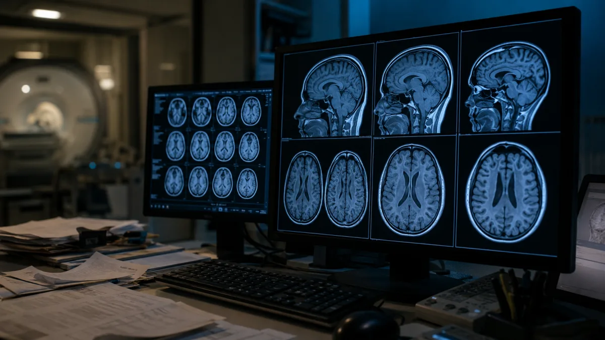
Glioma diagnosis begins with neuroimaging — typically MRI with and without contrast. Glioblastoma classically appears as a ring-enhancing lesion with central necrosis on contrast MRI. Definitive diagnosis requires tissue biopsy, which is analyzed both histologically and molecularly to determine IDH status, 1p/19q codeletion, MGMT promoter methylation, and other markers that inform prognosis and treatment selection.
For people diagnosed with glioblastoma, the standard initial treatment is the Stupp protocol: maximal safe surgical resection followed by concurrent radiation therapy and temozolomide (TMZ) chemotherapy, then adjuvant TMZ for six months. Tumor Treating Fields (TTFields), a device-based therapy that applies alternating electric fields to disrupt cell division, received FDA approval for newly diagnosed GBM following the EF-14 trial, which showed improved 5-year survival rates (13% versus 5% with TMZ alone). It is now included as a Category 1 option in NCCN guidelines for patients aged 70 and under.
For recurrent GBM, bevacizumab — an anti-VEGF antibody that reduces tumor vascularity — is used, primarily for its effect on symptoms and edema, though its impact on overall survival has been modest. IDH inhibitors (ivosidenib and enasidenib) are now used for IDH-mutant gliomas, targeting the molecular driver directly.
For lower-grade IDH-mutant gliomas, treatment is tailored to grade and extent of disease: watchful waiting for very slow-growing Grade 2 lesions in some patients, surgery plus radiation and chemotherapy (typically PCV — procarbazine, lomustine, vincristine — or temozolomide) for Grade 3 tumors.
Prognosis varies widely. GBM carries a median overall survival of approximately 14–15 months with standard therapy, with fewer than 10% of patients surviving five years. Lower-grade IDH-mutant gliomas carry substantially better outcomes: Grade 2 oligodendrogliomas may remain stable for many years, and patients with IDH-mutant tumors often survive five to ten years or more with treatment.
For anyone concerned about a family member's neurological changes or seeking broader context on related cancer prognoses, our article on metastatic bone cancer symptoms discusses how cancer spreading to other parts of the body presents differently from primary tumors.
Frequently Asked Questions
What is the most common cause of glioma?
No single cause accounts for most gliomas. The majority of cases arise from spontaneous DNA mutations in glial cells with no identifiable external trigger. The only confirmed environmental cause is prior exposure to therapeutic ionizing radiation. Inherited genetic syndromes such as neurofibromatosis type 1, Li-Fraumeni syndrome, and Lynch syndrome account for a small percentage of cases.
Are gliomas hereditary?
Most gliomas are not directly inherited. However, several rare hereditary syndromes — including neurofibromatosis type 1 and 2, Li-Fraumeni syndrome, Turcot syndrome, and Lynch syndrome — significantly increase the risk of developing a glioma. Having a first-degree relative (parent, sibling, child) with glioma is associated with roughly doubled risk, though the absolute probability remains low given how uncommon glioma is in the general population.
Do cell phones increase glioma risk?
Based on current evidence, cell phone use is not a confirmed cause of glioma. The IARC classified RF-EMF from mobile phones as "possibly carcinogenic" (Group 2B) in 2011, but subsequent large prospective studies — including the Million Women Study and the Danish Cohort Study — have not found an increased risk. A 2024 WHO-commissioned review concluded that mobile phones likely do not raise glioma risk. The question remains under investigation, but the evidence does not support a causal link as of 2025.
What is the difference between a glioma and a glioblastoma?
Glioma is the broader category — it covers all tumors arising from glial cells, including astrocytomas, oligodendrogliomas, and ependymomas across a range of grades. Glioblastoma (GBM) is a specific type of glioma: it is always IDH-wildtype, always WHO Grade 4, and the most aggressive primary brain tumor in adults. All glioblastomas are gliomas, but not all gliomas are glioblastomas.
What are the early warning signs of a glioma?
Early symptoms often include persistent and worsening headaches (typically worse in the morning), new-onset seizures in an adult who has never had them, gradual personality or mood changes, memory difficulties, vision problems such as blurred or double vision, and speech difficulties. Symptoms depend on the tumor's location. Because many of these signs overlap with common benign conditions, a glioma diagnosis typically requires brain imaging to identify.
When should I see a doctor about potential glioma symptoms?
See a doctor promptly if you experience: a new pattern of persistent, worsening headaches; a first-ever seizure; unexplained confusion or significant changes in memory or behavior; new visual disturbances; difficulty speaking or understanding language; or weakness or numbness in part of the body without an obvious injury-related cause. These symptoms rarely indicate a brain tumor, but they require evaluation. Earlier diagnosis generally allows more treatment options.
This article is for general informational purposes only and is not a substitute for professional medical advice. If you are experiencing persistent headaches, seizures, or neurological symptoms, please see a doctor promptly.
Gliomas remain among the most challenging brain cancers to treat, but the past decade has brought genuine advances: the molecular reclassification that now guides treatment decisions, FDA-approved tumor treating fields therapy, and IDH-targeted drugs for a subset of patients. Research continues on immunotherapy combinations and new surgical approaches. If you or someone you know has received a glioma diagnosis and wants to understand the outlook in more detail, our dedicated article on the prognosis for untreated glioblastoma provides specific survival data and context for treatment decisions. For authoritative medical information on glioma, the National Cancer Institute's brain tumor pages and Mayo Clinic's glioma overview are reliable starting points.

Linda Fehrman
Edits general wellness and relationship explainers. Health material is educational, avoids diagnosis and links to health-authority guidance.




Leave a reply
Replying to