
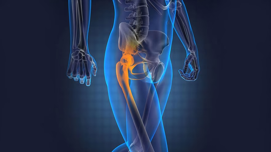
Bone is the third most common site of cancer metastasis in the body, after the lungs and liver. At any given time, an estimated 280,000 adults in the United States are living with cancer that has spread to their bones — a figure that dwarfs the roughly 3,450 new cases of primary bone cancer diagnosed each year, according to the National Cancer Institute. Understanding metastatic bone cancer symptoms early can be the difference between timely intervention and irreversible complications.
What is metastatic bone cancer and how is it different from primary bone cancer?
Primary bone cancer — osteosarcoma, chondrosarcoma, Ewing sarcoma — originates in the cells that make up bone tissue. It is rare, accounting for well under 1% of all new cancer diagnoses. Metastatic bone cancer is categorically different: it arises when malignant cells from a tumor elsewhere in the body break away, enter the bloodstream or lymphatic system, and take up residence in bone. The disease is still named and treated according to where it started. Breast cancer that spreads to the femur is breast cancer in the bone, not bone cancer — and that distinction shapes every treatment decision.
Once cancer cells arrive in bone, they hijack the organ's normal remodeling cycle. Healthy bone is in constant turnover, maintained by two opposing cell types: osteoblasts, which build new bone, and osteoclasts, which dissolve old or damaged bone. Tumor cells disrupt this balance, triggering one of two destructive patterns — or sometimes both — that erode structural integrity and generate the symptoms described below.
What does bone pain from metastases actually feel like?
Pain is the most frequently reported symptom of bone metastasis, and its character distinguishes it from the joint stiffness most people associate with arthritis. Arthritis pain typically responds to movement and eases after a joint warms up. Bone metastasis pain tends to do the opposite: it starts as a dull, deep ache and grows more severe at night, often waking the person from sleep. This nocturnal intensification happens partly because inflammatory mediators — prostaglandins and cytokines released by tumor cells — accumulate without the counterbalancing distractions of daily activity, and partly because recumbent positions shift mechanical load onto affected vertebrae and long bones.
Over time the pain transitions from intermittent to constant. Early on, weight-bearing aggravates it; later, even rest provides little relief. The pain can also radiate: a tumor in a thoracic vertebra may produce a band of discomfort around the chest wall that mimics pleurisy or a pulled muscle. A lesion near the hip joint may be felt in the groin or down the thigh. Unlike the gradual, symmetrical onset of osteoarthritis, bone metastasis pain is typically localized to one area and worsens progressively over weeks rather than years.
What causes pathological fractures in metastatic bone cancer?
A pathological fracture is a break that occurs in bone already weakened by disease — sometimes from nothing more forceful than rolling over in bed. Two distinct mechanisms drive this fragility, and understanding them explains why both feel so different structurally.
In osteolytic lesions, tumor cells — particularly those originating from breast cancer, kidney cancer, or multiple myeloma — flood the bone with signals that stimulate osteoclast activity far beyond normal levels. These overactive osteoclasts dissolve the mineralized bone matrix faster than osteoblasts can replace it, punching microscopic holes that merge into larger voids. Viewed on imaging, the cortex looks moth-eaten. The American Cancer Society reports that roughly 75% of bone metastases from breast cancer are osteolytic. The result is a bone that retains its shape on the outside but has been hollowed from within — structurally analogous to a wooden beam riddled with termite damage.
In osteoblastic lesions, seen most often with prostate cancer (70–85% of prostate bone metastases are osteoblastic according to Cleveland Clinic), tumor signals push osteoblasts into overdrive, depositing new bone at an abnormal rate. This new bone is structurally immature — dense in appearance on X-ray but laid down with disorganized collagen fibers, more like brittle calcium deposits than load-bearing cortical bone. Think of thin ice near a frozen riverbank: it looks solid but collapses under pressure. Both lesion types ultimately increase fracture risk, though through opposite cellular mechanisms.
The bones most commonly affected are the spine, pelvis, ribs, skull, upper femur, and humerus. A fracture in a weight-bearing bone causes sudden, severe pain and immediate loss of function.
What is hypercalcemia and why does it occur with bone metastases?

Bone is the body's largest calcium reservoir. When osteolytic metastases accelerate bone breakdown, calcium that was locked in the mineral matrix floods into the bloodstream. The result — hypercalcemia, defined as serum calcium above 10.5 mg/dL — affects an estimated 10–20% of people with advanced cancer at some point during their illness.
A second mechanism compounds this: many tumors — including certain brain malignancies (see What Are the Causes of Gliomas?) — secrete a molecule called parathyroid hormone-related protein (PTHrP), which mimics the hormone that normally raises blood calcium. PTHrP acts at distant sites — kidneys, intestines, bone — simultaneously reducing calcium excretion, increasing calcium absorption from food, and stimulating osteoclast activity. The two pathways reinforce each other in what researchers call a vicious cycle.
Symptoms of hypercalcemia are diffuse and easy to attribute to the cancer itself or to treatment side effects, which is why it often goes undetected until calcium levels become dangerously high. Early signs include nausea, constipation, loss of appetite, excessive thirst, and frequent urination. As calcium climbs further, confusion, muscle weakness, and irregular heart rhythms emerge. Severe hypercalcemia — above 14 mg/dL — can lead to coma. Any cancer patient experiencing unexplained confusion alongside bone pain should have calcium levels checked promptly.
What are the other symptoms of metastatic bone cancer?
Beyond the cardinal triad of pain, fractures, and hypercalcemia, bone metastasis produces a cluster of symptoms that reflect either the systemic burden of advanced cancer or the direct effects of tumor infiltration in specific locations.
Fatigue and weakness are near-universal in metastatic disease. For older patients in particular, maintaining motivation during prolonged treatment can be challenging — strategies from How to Motivate the Elderly may offer practical guidance for caregivers. Cancer-related fatigue differs from ordinary tiredness in that it does not resolve with rest. It stems from multiple overlapping causes: anemia from bone marrow infiltration reducing red blood cell production, inflammatory cytokines from tumor cells altering hypothalamic function, disrupted sleep from pain, and the metabolic cost of an immune system in constant activation.
Anemia deserves particular attention when metastases involve the axial skeleton — the vertebrae, pelvis, and ribs — because these bones contain the greatest volume of active bone marrow. When tumor cells displace marrow, red blood cell production falls. Anemia manifests as pallor, fatigue, exertional shortness of breath, rapid heartbeat, and dizziness. These symptoms belong to the anemia, not directly to the bone disease, but they signal marrow involvement and should prompt imaging.
Swelling may appear as a visible or palpable mass over an affected bone, particularly when a lesion lies close to the skin surface or expands into adjacent soft tissue. In the spine, tumor growth into the epidural space is the mechanism behind one of the most feared complications: spinal cord compression.
Fever occurs in some patients as part of a paraneoplastic response — tumors secrete interleukins and other pyrogens that raise core body temperature. It tends to be low-grade (99–101°F), intermittent, and accompanied by night sweats. A fever in someone with known cancer always warrants evaluation, as infection — especially in immunocompromised patients — must be excluded.
Spinal cord compression is an oncological emergency. When vertebral metastases collapse or tumor extends into the spinal canal, pressure on the cord produces back pain radiating down one or both legs, progressive leg weakness, and eventually loss of bladder or bowel control. Numbness or tingling in the legs, feet, or lower abdomen requires immediate emergency evaluation — permanent paralysis can develop within hours if compression is not relieved. Call 911 or go to the nearest emergency room if these symptoms appear.
Which cancers most commonly spread to bone?
While virtually any cancer can metastasize to bone, a handful of primary tumors account for the vast majority of cases. Prostate and breast cancers top the list: up to 65–80% of patients with advanced prostate or breast cancer will develop bone metastases during their disease course, according to published epidemiological data. Lung cancer follows, with bone involvement in roughly 40–50% of metastatic cases, though survival after lung-to-bone metastasis is markedly shorter — approximately six to seven months, compared with 53 months for prostate-to-bone metastasis, per Cleveland Clinic data.
Kidney (renal cell) carcinoma and thyroid cancer also have a strong predilection for bone, and their lesions tend to be highly osteolytic with a rich blood supply, making them prone to bleeding and difficult to treat surgically. Multiple myeloma, though technically a blood cancer arising in bone marrow plasma cells, produces bone lesions that are mechanically and clinically indistinguishable from osteolytic metastases — and is the most common malignancy to involve the skeleton overall.
The bones at greatest risk reflect skeletal blood flow: red marrow-rich sites in the axial skeleton (vertebral column, pelvis, ribs, sternum) are far more commonly seeded than the appendicular skeleton. This is why back pain in a patient with a known cancer history should never be assumed to be simple degenerative disease without imaging.
How is metastatic bone cancer diagnosed?
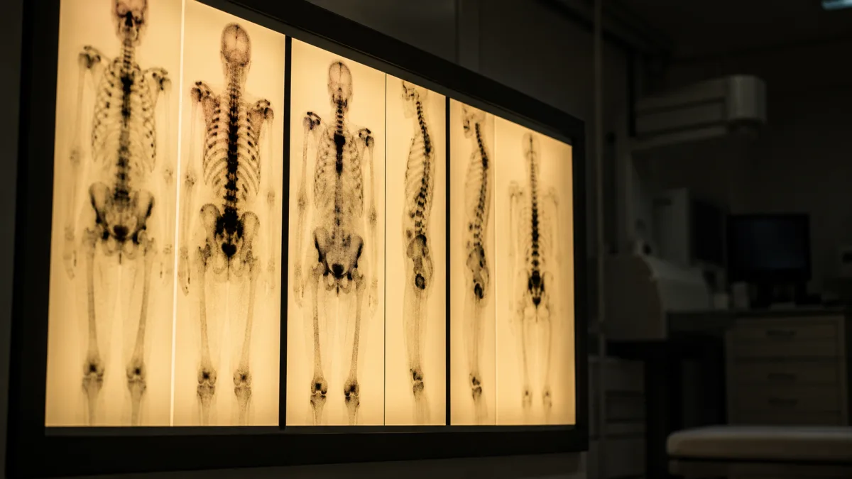
Diagnosis typically begins with plain X-rays, which can reveal osteolytic holes or osteoblastic densities — but X-rays miss early or small lesions, and up to 50% of trabecular bone must be destroyed before a lesion appears on a standard film. A technetium-99 bone scan (scintigraphy) offers whole-skeleton coverage and detects areas of abnormal bone turnover earlier than X-ray, but it lacks anatomical detail and can produce false positives from fractures, infections, or arthritis.
MRI has emerged as the most sensitive modality for detecting bone marrow involvement and soft-tissue extension. It uses no ionizing radiation and provides superior contrast between healthy marrow and tumor infiltration — particularly valuable for evaluating spinal lesions and risk of cord compression. CT scanning complements MRI by showing the state of the cortical bone in fine cross-sectional detail, which is critical for fracture risk assessment before weight-bearing rehabilitation.
PET scanning, typically using fluorodeoxyglucose (FDG), detects metabolically active lesions based on the preferentially high glucose uptake of cancer cells. It is especially sensitive for osteolytic metastases and can reveal disease before structural bone changes appear. Blood tests — measuring alkaline phosphatase, lactate dehydrogenase, and serum calcium — provide supporting evidence and help monitor treatment response. A bone biopsy may be required when a lesion appears in isolation and the primary cancer is unknown or when the imaging picture is ambiguous. The National Cancer Institute recommends biopsy be performed by an orthopedic oncologist, as incision placement affects subsequent surgical options.
What treatments are available for metastatic bone cancer?
Treatment goals for bone metastasis are predominantly palliative — reducing pain, preventing skeletal complications, and maintaining quality of life — rather than curative. A combination of local and systemic approaches is typically used.
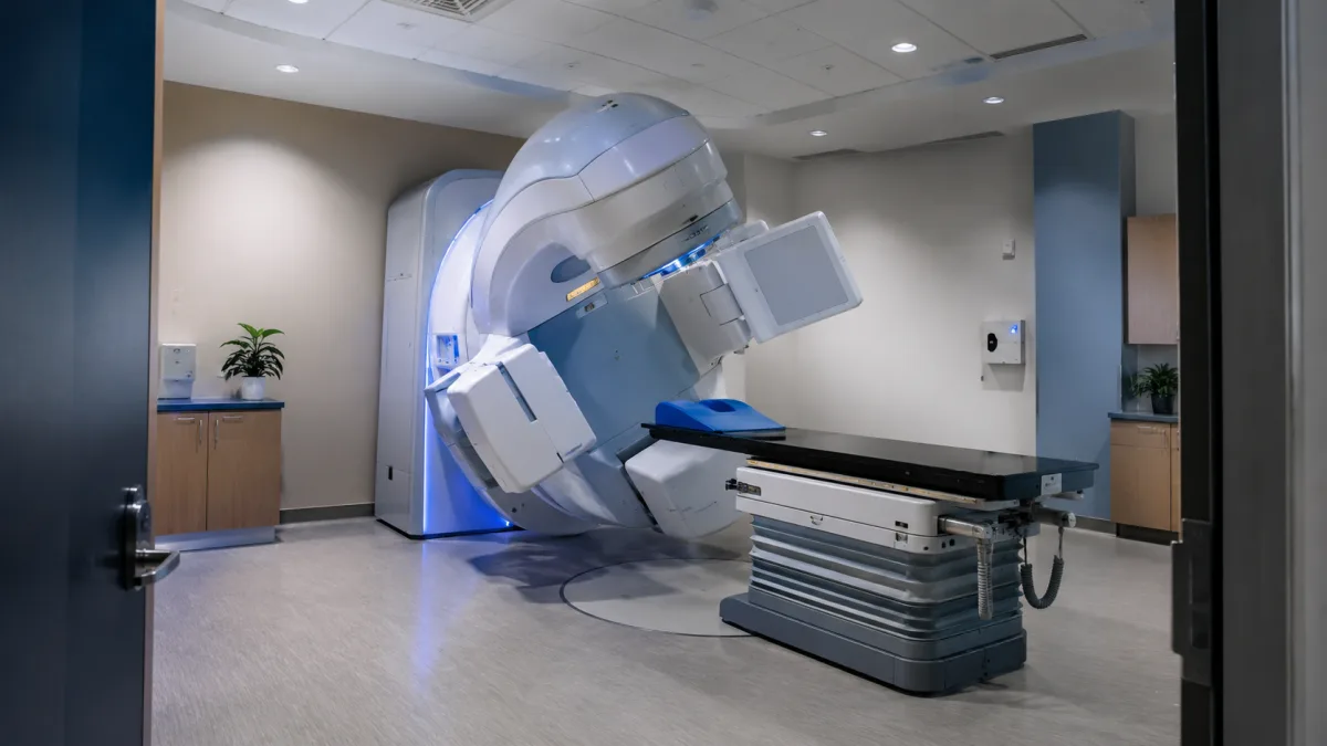
Radiation therapy directed at a painful bone lesion reduces pain in approximately 60–80% of patients, often within two to three weeks. For spinal cord compression, it is combined with high-dose corticosteroids to reduce edema around the cord rapidly. Stereotactic body radiotherapy (SBRT) can deliver high-dose, precisely targeted radiation to vertebral or other discrete metastases with minimal surrounding tissue damage.
Bisphosphonates — pamidronate (Aredia) and zoledronic acid (Zometa) — work by inhibiting osteoclast function, slowing the bone resorption that drives osteolytic lesions. Intravenous zoledronic acid is the most widely used agent and reduces the incidence of fractures, the need for radiation, and hypercalcemia events. Denosumab (Xgeva) achieves a similar effect through a different molecular target — blocking RANKL, the signaling protein that promotes osteoclast maturation — and has shown superiority over zoledronic acid in some trials for preventing skeletal-related events.
Systemic therapies targeting the primary cancer — chemotherapy, hormone therapy for breast or prostate cancer, targeted agents, or immunotherapy — can simultaneously shrink bone metastases by attacking tumor cells wherever they reside. Surgery is reserved for impending or completed pathological fractures, typically involving long bones, where internal fixation or joint replacement can restore function. Kyphoplasty — injecting bone cement into fractured vertebrae under image guidance — provides rapid pain relief and structural stabilization without open surgery.
Clinical trials are actively testing novel approaches, including radiopharmaceuticals that home in on bone-seeking tumors, and combinations of denosumab with immune checkpoint inhibitors. Patients should ask their oncologist whether any trials are appropriate for their specific cancer type and stage.
Frequently Asked Questions
Can metastatic bone cancer be cured?
In most cases, bone metastasis is not curable — it indicates that cancer has spread beyond its primary site and is considered stage IV disease. Treatment focuses on controlling the disease, relieving symptoms, and preserving function. That said, survival times vary enormously by cancer type: prostate cancer with bone metastases has a median survival exceeding four years, while lung cancer with bone involvement carries a median survival closer to six months. For context on how different advanced brain cancers compare, see The Prognosis for Untreated Glioblastoma.
Is all bone pain in a cancer patient caused by metastasis?
No. Cancer patients can have unrelated musculoskeletal conditions — degenerative disc disease, osteoporosis fractures, or treatment-related bone loss from steroids or aromatase inhibitors — that produce bone pain. This is why imaging and sometimes biopsy are essential. Any new, unexplained bone pain in a person with a cancer history warrants prompt evaluation.
What is a skeletal-related event?
Clinicians use the term "skeletal-related event" (SRE) to describe the major complications of bone metastasis: pathological fracture, spinal cord compression, need for radiation to bone, and surgery to bone. SREs are the primary endpoint in clinical trials testing bone-protective drugs like bisphosphonates and denosumab, because they represent measurable, significant deteriorations in a patient's functional status.
How is bone metastasis pain managed day to day?
Pain management typically follows a stepwise approach: non-opioid analgesics (NSAIDs, acetaminophen) for mild pain, progressing to short-acting then long-acting opioids for moderate to severe pain. Bone metastasis pain often responds better when opioids are combined with NSAIDs because the inflammatory component of bone pain (driven by prostaglandin release from tumor cells) responds to anti-inflammatory drugs in ways that opioids alone do not fully address. Palliative care teams, working alongside the oncologist, specialize in achieving adequate pain control without compromising alertness or quality of life.
Should I limit physical activity if I have bone metastases?
Not necessarily — but activity should be tailored carefully. Bones with high-risk osteolytic lesions, particularly in weight-bearing locations, may require activity restrictions to reduce fracture risk. A physical therapist experienced in oncology rehabilitation can assess each patient individually and design a safe exercise program. Immobility itself carries risks: muscle wasting, blood clots, and worsening fatigue all accelerate with inactivity. Ask your oncologist and a physical therapist to collaborate on what is safe for your specific lesion locations and severity.
When is it an emergency?
Seek emergency care immediately if you experience sudden numbness or tingling in the legs, feet, or lower abdomen; inability to move the legs normally; loss of bladder or bowel control; or inability to urinate. These are signs of spinal cord compression. Untreated, spinal cord compression can cause permanent paralysis within hours. Similarly, severe hypercalcemia — marked by profound confusion, lethargy, or loss of consciousness — requires emergency hospitalization.
This article is for general informational purposes only and does not constitute medical advice. If you or a loved one are experiencing these symptoms, consult an oncologist or your primary care physician promptly.
The most actionable guidance for anyone with a known cancer history and new bone pain is straightforward: call your oncologist before assuming it is muscular or arthritic. Early imaging changes treatment options — a lesion caught before pathological fracture is far more amenable to stabilization than one discovered after the bone has already given way. Do not wait for the pain to become unbearable. Early reporting to your care team is the single most protective step you can take.

Tory Stearns
Edits practical household, travel and lifestyle explainers. Claims that can change are linked to current primary or subject-authority sources.




Leave a reply
Replying to